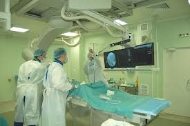
Vascular angiography
Vascular angiography: contraindications, preparation, performance, aftereffects
Angiography is an X-ray examination of blood vessels. It may be used in radiography, hybrid surgical, computed tomography. This modality enables to study the functional state of blood vessels, blood flow, and the extent of abnormal spread.
The ordinary X-ray films do not allow to see arteries and veins, as the vessels are able to absorb X-rays, just as the adjacent tissues are.

What diseases can be detected?
Angiography can detect the following abnormalities of blood vessels and organs they supply with blood:
- Impaired patency as a result of thrombosis or atherosclerosis;
- Aneurysms;
- Malformation;
- Defects and disorders of the internals;
- Tumors;
- Vascular heart diseases.
Angiography also enables to conduct interventional curative procedures: embolization, chemoembolization of tumor vessels.
Contraindications
Angiography is contraindicated with:
- acute diseases of infectious and inflammatory nature;
- mental diseases;
- renal and hepatic failure;
- heart diseases;
- hemostasis disorders;
- venereal diseases;
- thyroid dysfunctions;
- allergy to iodine and its preparations;
- anesthesia allergy;
- patient’s grave condition.
Preparation
To conduct the procedure, patient’s consent is needed.
Preliminary ECG and photoroentgenography are performed.
Patients are advised to give up spirits 14 days prior to angiography.
To protect kidneys from iodine exposure, hydration is carried out, i.e. the body is saturated with liquid: it dilutes the radiopaque medium and facilitates its quick washout.
No food or drink are taken before the procedure (about 4 hours before its start).
Hair is removed in the puncture area.
All metal decorations and other things are taken off.
Performance
The patient lies down on a special angiography table. He/she is fixed and connected to cardiomonitor.
Intravenous catheter is inserted. It is used for premedication ¬ introduction of the following drugs:
- antihistamines to prevent possible allergic reaction;
- tranquilizers;
- analgetics.
Angiography may be performed by means of puncture (paracentesis) or catheterization¬ insertion of catheter for contrast medium into a blood vessel. Inmost of the cases, femoral artery is catheterized.
Following local anesthesia, a 3-4 mm incision is made in the skin. A puncture is made on the artery with a special wide-lumen needle. The needle is needed for the subsequent introduction of a mental conductor being then moved to the required level. The conductor itself is removed. The operations inside the vessel are roentgenotelevision-guided.
The contrast medium is introduced via the installed catheter. Beginning with this moment, preliminarily programmed X-ray velocity filming is made.
The films having been analysed, additional films may be made, if needed.
The catheter is removed, and sterile dressing is applied to the puncture area. It must remain there for 24 hours.
Current angiography
Only subtraction digital angiography is employed in current medicine. Its essence is in contrast-enhanced examination of vessels and subsequent computer-based processing.
The advantages of the modality:
- It enables to obtain better-quality films allowing to discern separate vessels out of the whole pattern;
- The opportunity to reduce the dose of the radiopaque medium introduced;
- The introduction of the contrast medium can be performed without catheterization; owing to this, traumatizing caused by the procedure is reduced.
Post-angiography period
A 24-hour bed rest is indicated for the patient subjected to angiography. He/she needs to be under doctor’s care. The puncture site is recurrently inspected, and body temperature is measured. For rapid excretion of iodine and other drugs from the body, patients are advised to consume large amounts of liquid.
Possible aftereffects
The procedure may cause some complications, such as:
- emergence of hematoma in the puncture area;
- thromb obstruction of artery lumen (thromboembolism) very rare occurrence
In Minsk, this procedure is provided at N. N. Alexandrov National Cancer Centre, the leading oncological institution of Belarus.


