Ultrasonic diagnosis
US (ultrasonic diagnosis) is a non-invasive study of the body or the inner structure of various objects and processes ongoing in them, by means of ultrasound waves.
Its main advantages are the absence of contraindications and significant adverse effect on the organ or object under investigation. This modality is used for primary and subsequent diagnosis of diseases, assessment of the disease course, detection of recurrence at early stages, determining the magnitude of surgical intervention and diagnosis of possible complications of the treatment.
US advantages
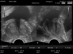
Presently, various techniques are used for ultrasonography in oncology: B-mode, Doppler ultrasonography, elastography, M-mode and others. US studies can be conducted either transcutaneously or with special cavitary (endovaginal, endorectal, transesophageal, intravascular) and intraoperative gauges of different form and configuration.
The minimal resolving power of current ultrasonic scanners reaches 1-2 mm, enabling to visualize objects of the sizes mentioned as separate structures. This has become possible owing to high-frequency and broad-band gauges.
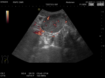
Doppler techniques such as energy coloured mapping, 3D panoramic reconstruction of vessels are capable of assessing vascular architectonics in the tumor area.


